
男女共同参画学協会連絡会
支援企業による広告記事
- ソーラボジャパン株式会社
- CAD を使って光学装置を設計してみよう
- 「生物物理」2025年10月号
- ソーラボジャパン株式会社
- 次世代2光子顕微鏡―小型化がもたらす新たな可能性
- 「生物物理」2024年10月号
- ソーラボジャパン株式会社
- サイエンティフィックカメラと周辺機器の同期
- 「生物物理」2023年10月号
- ソーラボジャパン株式会社
- 顕微鏡のリノベーション ~ 顕微鏡ポートを活用した光学系の導入
- 「生物物理」2022年12月号
「Biophysics and Physicobiology」に Takahiro Fujiwara, Akihiro Kusumi による "Development of ultrafast single fluorescent-molecule imaging and its application to unravel plasma membrane structure and function in live cells" をJ-STAGEの早期公開版として掲載
2026年05月26日 学会誌
日本生物物理学会欧文誌[Biophysics and Physicobiology]に以下の論文が早期公開されました。
Takahiro Fujiwara, Akihiro Kusumi
"Development of ultrafast single fluorescent-molecule imaging and its application to unravel plasma membrane structure and function in live cells"
URL:https://doi.org/10.2142/biophysico.bppb-v23.0018
- Abstract
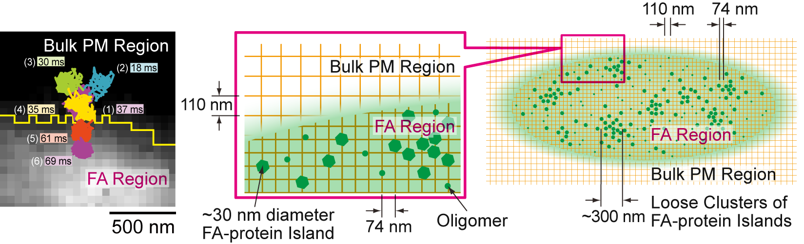
- Single-molecule imaging in live cells now plays key roles in elucidating molecular dynamics and interactions. However, the imaging time resolution has remained limited, despite its critical importance for precisely capturing molecular events in cells, in contrast with major spatial resolution advances in fluorescence microscopy. To address this issue, we developed an ultrafast camera system that achieves the highest time resolutions reported to date for single fluorescent-molecule imaging and tracking (SFM-IT), reaching fluorophore photophysics-limited (photon-limited) frame times of 33 and 100 μs with single-molecule localization precisions of 34 and 20 nm, respectively, for Cy3, the optimal fluorophore identified. Using this system, we directly detected the hop diffusion of membrane molecules in the plasma membrane, confirming its compartmentalization. Thus, ultrafast SFM-IT has helped to elucidate the principles governing the plasma membrane organization and molecular dynamics. Building on this platform, we further established ultrafast, live-cell, two-color single-molecule localization microscopy (SMLM). This method reduced the data acquisition time by ≈30-fold relative to standard methods, while simultaneously enabling much larger view-fields, with localization precisions of 29 and 19 nm for PALM and dSTORM, respectively. Combining ultrafast SMLM with ultrafast SFM-IT revealed the mesoscopic dynamical organization of focal adhesions (FAs), termed an archipelago architecture, showing that FAs consist of ≈30-nm-diameter FA-protein islands loosely clustered into ≈300-nm-diameter functional units embedded in the compartmentalized fluid membrane (74 nm inside vs. 110 nm outside the FA). Ultrafast SFM-IT and ultrafast SMLM techniques open previously inaccessible spatiotemporal regimes for biophysical cell biology research.
URL: https://doi.org/10.2142/biophysico.bppb-v23.0018