

Ranking 1 Access:52441

2013.07.03
Species name: Amoeba proteus
University of Hyogo, Graduate School of Life Science Yukinori Nishigami
Amoeba proteus exhibit active cell locomotion. During locomotion, cytoplasm actively flows toword direction of locomotion.
Ranking 2 Access:38466

2014.03.13
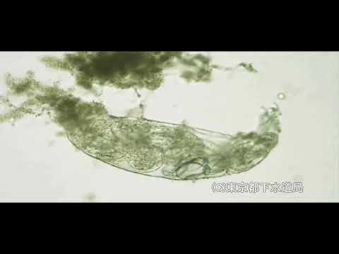
Species name: Euglena
Department of Biology, Faculty of Science, Kobe University Masashi HAYAKAWA
Euglenoid movement of Euglena
Ranking 3 Access:34894
2017.08.29
Species name: Macrobiotus sp.
Tokyo Metropolitan Government Bureau Swerage,
The size of Macrobiotus is about 0.2 – 1.0 mm. The body is covered with a thin chitin film, with spiny bristles, armor plates. There are four pairs of footsteps, with nails at the tip. Macrobiotus have fourth pair of legs with nails at the tip. They usually live in soil. The form that I'm slow in action and walk slowly is similar to a bear, so it's called a bear bug. The tooth needle taken out of the mouth is stuck into food, and it's crowded, and a pharynx, to work, more, I suck at contents. Slowly walking figure resembles a bear so it is called water bears.
Ranking 4 Access:33127

2013.09.03
Species name: Paramecium caudatum
Faculty of Science, Yamaguchi University Yoshiaki Iwadate
It is not well known that Paramecium cells discharge missiles named "trichocyst" when they are attacked by a predator. Trichocyst discharge and ciliary reversal in Paramecium cells are regulated by the intracellular calcium concentration. We injected caged Calcium into P. caudatum cells and applied ultraviolet (UV) light to the cell for 125ms. This did not induce trichocyst discharge but did induce ciliary reversal . A re-application of UV for 125 ms triggered trichocyst discharge. The threshold of intracellular calcium for trichocyst discharge is higher than those for ciliary reversal.
Iwadate et al., Protoplasma 206, 11-19, 1999
Iwadate and Nakaoka, Cell Calcium 44, 169-179, 2008
Ranking 5 Access:14432

2014.07.19
Species name: Rabbit
Osaka City Univ Eisaku KATAYAMA
The first part of the movie indicates the movement of individual myosin-head (crossbridge), based on conventional "Tilting-Leverarm Hypothesis". Such movement was proposed from the characteristic features of the atomic models of myosin-S1 in the absence and the presence of ATP, together with the well-known experimental evidence that "the motor-domain does not appreciably rotate during the Power-Stroke". Hence, this hypothesis claims that the Power-Stroke is essentially the transition between strongly actin-bound rigor-structure (1DFK: lever-arm is extended) and ATP-bound kinked structure (1DFL). If the motor-domain is immobilized on actin, the lever-arm moiety should swing along the actin filament, The latter half of the movie exhibits the revised crossbridge-cycle we have proposed according to our direct observation of in vitro sliding actomyosin by Quick-Freeze Deep-Etch Replica Electron Microscopy (Ref. 1, 2). We noticed that the actual images of actin-sliding myosin cannot be explained by the conventional hypothesis as above, suggesting the presence of a new conformer whose crystal structure is not yet reported. After extensive search, we finally found that SH1-SH2 crosslinked myosin could be a good candidate of the new conformer whose lever-arm bends to the opposite side of ATP-bound kinked structure (Ref.3-5). Since we could successfully reconstruct its low-resolution 3-D model by a new version of single-particle-analysis (Ref. 5). Taking the results of time-resolved chemical crosslinking into consideration, we revised the scheme of crossbridge-cycle including the new conformer (Ref.5). .The conformational change shown in the movie is compatible with all the images we actually observed under in vitro actin-sliding conditions. [References] 1. Katayama E. The effects of various nucleotides on the structure of actin-attached myosin subfragment-1 studied by quick-freeze deep-etch electron microscopy. J Biochem. 1989 Nov;106(5):751-70. 2: Katayama E. Quick-freeze deep-etch electron microscopy of the actin-heavy meromyosin complex during the in vitro motility assay. J Mol Biol. 1998 May 1;278(2):349-67. 3: Katayama E, Ohmori G, Baba N. Three-dimensional image analysis of myosin head in function as captured by quick-freeze deep-etch replica electron microscopy. Adv Exp Med Biol. 1998;453:37-45. 4: Katayama E, Ichise N, Yaeguchi N, Yoshizawa T, Maruta S, Baba N. Three-dimensional structural analysis of individual myosin heads under various functional states. Adv Exp Med Biol. 2003;538:295-304. 5: Kimori Y, Baba N, Katayama E. Novel configuration of a myosin II transient intermediate analogue revealed by quick-freeze deep-etch replica electron microscopy. Biochem J. 2013 Feb 15;450(1):23-35. 6. Andreev OA, Reshetnyak YK. Mechanism of formation of actomyosin interface. J Mol Biol. 2007 Jan 19;365(3):551-4.
Ranking 6 Access:11955

2015.07.10
Species name: Mus musculus
Hamamatsu Univ Sch Med Koji Ikegami
The fluid flow is driven by the well coordinated ciliary beating. The flow speed is around 25 micrometer per sec, which is equal to ~1.5 mm/min or 1 cm/6-7 min. Tiny particles or pathogens such as viruses are removed from the respiratory organ by this flow.
Ranking 7 Access:10908

2016.12.15
Species name: Amphisiella
AL-Museum AL-Museum
This ciliate is busily foraging back and forth, changing directions and passing between particles. There are cirri at the front and rear of its slender body, and the cirri at the front are especially active.
Ranking 8 Access:10366

2015.10.30
Species name: Mycoplasma mobile
Osaka City Univ Makoto MIYATA, Tasuku HAMAGUCHI
supplemented to a scientific paper Prospects for the gliding mechanism of Mycoplasma mobile Makoto Miyata, Tasuku Hamaguchi (Osaka City University) doi:10.1016/j.mib.2015.08.010 end user license: CC BY-NC-ND 4.0
Ranking 9 Access:8877

2015.06.25
Faculty of Life and Environmental Scienses, Prefectural University of Hiroshima Shin-ichi Aizawa
Bacterial flagellar filament has polymorphic transition ability, which can switch between a set of helical forms (straight, Normal coiled and curly) in response to flagellar motor rotation, pH, salinity, and temperature changes.
Ranking 10 Access:7946

2015.06.25
Faculty of Life and Environmental Scienses, Prefectural University of Hiroshima Shin‒ichi Aizawa
Bacterial flagellar filament has polymorphic transition ability, which can switch between a set of helical forms (straight, Normal coiled and curly) in response to flagellar motor rotation, pH, salinity, and temperature changes.
Ranking 11 Access:7540

2014.10.08
School of Molecular Bioscience, University of Sydney, Sydney, New South Wales, Australia Professor Timothy P. Newsome
HeLa cells were infected with B5R-YFP (green), transfected with pE/L Lifeact-mRFP (red) and imaged for 80 seconds at 6–9 hours post infection. A virus particle is seen co-localizing with F-actin tail of approximately 3–4 µm.
Ranking 12 Access:7259

2017.08.09
Waseda Univerisity Shin'ichi ISHIWATA
Standard SPOC
Ranking 13 Access:7203

2016.11.14
Species name: Paenibacillus sp.
Graduate School of Biological Sciences, Nara Institute of Science & Technology Kazuo Kobayashi
Time-lapse analysis of colony formation. One microliter of Paenibacillus sp. suspension was spotted onto a 1.5% agar plate, which was then incubated at 37°C. The process of colony formation was analyzed by time-lapse light microscopy. Time-lapse images were collected every 1 min for 16 h after inoculation. Playback speed, ×1,440. Scale bar, 2 mm.
Genetic Analysis of Collective Motility of Paenibacillus sp. NAIST15-1
Ranking 14 Access:7039

2017.08.02
Waseda Univerisity Shin'ichi ISHIWATA
SPOC(Myofibril in auxotonic condition)
Ranking 15 Access:7025

2015.05.25
Species name: Amoeba proteus
National Institute for Basic Biology TANIGUCHI, Atsushi
Locomotion of Amoeba
Ranking 16 Access:6910

2015.06.25
Faculty of Life and Environmental Scienses, Prefectural University of Hiroshima Shin‒ichi Aizawa
Bacterial flagellar filament has polymorphic transition ability, which can switch between a set of helical forms (straight, Normal coiled and curly) in response to flagellar motor rotation, pH, salinity, and temperature changes.
Ranking 17 Access:6904

2017.08.09
Waseda Univerisity Shin'ichi ISHIWATA
Fig1bSide_Branching by Arp2/3complex with VCA
Ranking 18 Access:6898

2017.08.02
Waseda Univerisity Shin'ichi ISHIWATA
Super helix formation of actin filaments.
Right-handed rotation of an actin filament in an in vitro motile system (Nature, 1993, 361:261-71)
Ranking 19 Access:6866

2017.08.02
Waseda Univerisity Shin'ichi ISHIWATA
rabbit SPOC
Ranking 20 Access:6814

2016.11.14
Species name: Paenibacillus sp.
Graduate School of Biological Sciences, Nara Institute of Science & Technology Kazuo Kobayashi
Cellular behavior of the wild-type strain at the leading edge zones of swarming colonies on 0.6% agar plates. The wild-type strain was spotted onto the center of 0.6% agar plates and incubated at 37°C for 6 h. Coverslips were placed directly on the surface of the leading edge zones of the colonies and cell morphology observed under a video light microscope. These movie is real time. Scale bar, 20 μm.
Genetic Analysis of Collective Motility of Paenibacillus sp. NAIST15-1
Ranking 21 Access:6585

2017.08.09
Waseda Univerisity Shin'ichi ISHIWATA
Fig1eEnd_Branching by Arp2/3complex with VCA
Ranking 22 Access:6522

2016.11.14
Species name: Paenibacillus sp.
Graduate School of Biological Sciences, Nara Institute of Science & Technology Kazuo Kobayashi
Small moving clusters of wild-type strain cells on a 1.5% agar plate. The wild-type strain was spotted on a 1.5% agar plate and incubated at 37°C for 6 h. Coverslips were placed directly onto the surface of the leading edge zones of the colonies and cell morphology observed under a video light microscope. The movie is real time. Scale bar, 20 μm.
Genetic Analysis of Collective Motility of Paenibacillus sp. NAIST15-1
Ranking 23 Access:6512

2016.11.14
Species name: Paenibacillus sp.
Graduate School of Biological Sciences, Nara Institute of Science & Technology Kazuo Kobayashi
Large moving clusters of wild-type strain cells on a 1.5% agar plate. The experiment was done as described in "Motility of Paenibacillus sp. NAIST15-1 (1)" Movie. The movie is real time. Scale bar, 20 μm.
Genetic Analysis of Collective Motility of Paenibacillus sp. NAIST15-1
Ranking 24 Access:6444

2017.08.02
Waseda Univerisity Shin'ichi ISHIWATA
rat SPOC
Ranking 25 Access:6443

2016.02.08
Osaka City University Hamaguchi Tasuku
Quick freezing made by Blender (free- software).
Ranking 26 Access:6420

2016.11.14
Species name: Paenibacillus sp.
Graduate School of Biological Sciences, Nara Institute of Science & Technology Kazuo Kobayashi
The cmoA mutant retains functional flagella. cmoA mutant cells were spread over the surface of a 1.5% agar plate and incubated at 37°C for 5 h. Two hundred microliters of water were poured onto the colonies and cellular behavior was immediately observed under a video light microscope. The movie is real time. Scale bar, 20 μm.
Genetic Analysis of Collective Motility of Paenibacillus sp. NAIST15-1
Ranking 27 Access:6402

2014.08.21
University of California, San Francisco Professor Ronald D. Vale
Time-lapse observation of GFP-tubulin in wild-type S2 cells during bipolar spindle formation. Images are taken every 10 sec using spinning-disk confocal microscopy.
Ranking 28 Access:6203

2017.08.02
Waseda Univerisity Shin'ichi ISHIWATA
Actin Polymerization Triggered by Arp2/3complex.
Ranking 29 Access:6103

2017.08.09
Waseda Univerisity Shin'ichi ISHIWATA
Fig1gEnd_Coupling by Arp2/3complex with VCA
Ranking 30 Access:5994

2014.10.14
Instituto de Tecnologia Quı´mica e Biolo´ gica, Universidade Nova de Lisboa, Oeiras, Portugal Professor Claudio M. Soares
MalK dimer interface dissociation in the ADP-bound state. The dimer opening occurred in one of the ten replicates after ~37 ns of MD simulation. At the beginning of the simulation (t=0 ns), both binding sites are closed and the two ADP molecules are bound at the interface between monomers. At the end of the simulation (t=50 ns), binding site 2 is completely separated and the nucleotide is only bound to the P-loop residues.